Scientists found that three conserved regions play important roles in the enzymatic activity and function of P33
Sulfhydryl oxidase catalyzes disulfide bond formation of substrate proteins. P33, a baculovirus-encoded sulfhydryl oxidase, is different from other cellular and viral sulfhydryl oxidases, bearing unique features in tertiary and quaternary structure organizations.
To better understand the structural and functional relationship of P33, a series of point mutations in three conserved regions were generated and their biological functions were studied by the research group led by Prof. HU Zhihong and the research group led by Prof. GONG Peng from Wuhan Institute of Virology of the Chinese Academy of Sciences. The sulfhydryl oxidase activities of these mutants were biochemically tested in vitro. Correspondingly, recombinant viruses were constructed to study the biological function of the mutants in vivo. Crystal structures of wild-type (wt) and representative P33 mutants were also resolved to aid the analyses of the functional data.
Their results showed that the three conserved regions, i.e. the active site, dimer interface and the R127-E183 salt bridge, are key regions of P33 and play significant roles in virus morphogenesis and oral infectivity.
The results have been published in Journal of Virology entitled "Three conserved regions in baculovirus sulfhydryl oxidase, P33, are critical for enzymatic activity and function ".
This research was supported by the grants from the National Natural Science Foundation of China, the Strategic Priority Research Program of the Chinese Academy of Sciences, the National Key Basic Research Program of China, the Keyresearch projects of frontier science, Chinese academy of sciences, the National Key R&D Program of China and the Virology Key Frontier Science Program of State Key Laboratory of Virology.
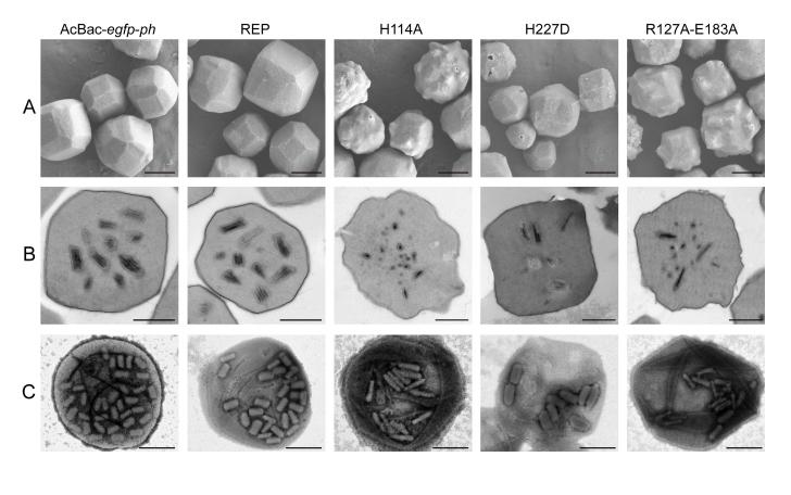
Fig.1. Electron microscopy (EM) analysis of OBs purified from infected larvae. (A) OBs purified from the infected larvae were visualized by SEM to observe the surface morphology. Image by HU Zhihong
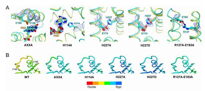
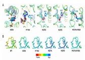
Fig.2. Crystallographic characterization of P33 mutants and a structure-based flexibility analysis of wt and mutant P33 constructs. Image by HU Zhihong
Contact:
HU Zhihong
Email: huzh@wh.iov.cn
Wuhan Institute of Virology, Chinese Academy of Sciences, Wuhan 430071, China (http://english.whiov.cas.cn/)