Scientists map a single-cell atlas for Aedes aegypti's midgut
Date:07-06-2024 | 【Print】 【close】
To address this issue, the research groups led by YUAN Zhiming and XIA Han from Wuhan Institute of Virology of the Chinese Academy of Sciences, and CUI Yingjun from Yale School of Medicine have successfully constructed a single-cell level midgut atlas of adult female Aedes aegypti mosquitoes, which published in Scientific Data entitled “A cell atlas of the adult female Aedes aegypti midgut revealed by single-cell RNA sequencing” (https://www.nature.com/articles/s41597-024-03432-8).
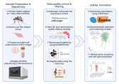
In this study, the midguts of 7-day-old adult female Aedes aegypti fed only glucose were dissected, collected, and prepared into single-cell suspensions by enzymatic dissociation and density gradient centrifugation protocols. The suspensions were then used for the single-cell RNA sequencing with the 10X Genomics platform (Fig. 1), and the cell atlas of mosquito midgut was characterized by the downstream analysis. Six types of intestinal cells including intestinal stem cells/enteroblasts (ISC/EB), cardia cells (Cardia), enterocytes (EC), enterocyte-like cells (EC-like), enteroendocrine cells (EE) and visceral muscle (VM) were obtained by clustering and annotating the transcriptomic features of single cells (Fig. 2). Additionally, marker genes for different cell types were also identified (Fig. 2). This study will lay the foundation for further research to analyze the patterns of viral infection and transmission with mosquitoes at the single-cell level.
Mr. WANG Shunlong and Dr. HUANG Ying are the co-first authors, and Prof. YUAN Zhiming, Prof. XIA Han, and Associate research scientist CUI Yingjun are the co-corresponding authors. This work was supported by grants from the National Key Research and Development Program of China and the National Natural Sciences Foundation of China.

Fig 1. Flow diagram of the scRNA-seq and data analysis
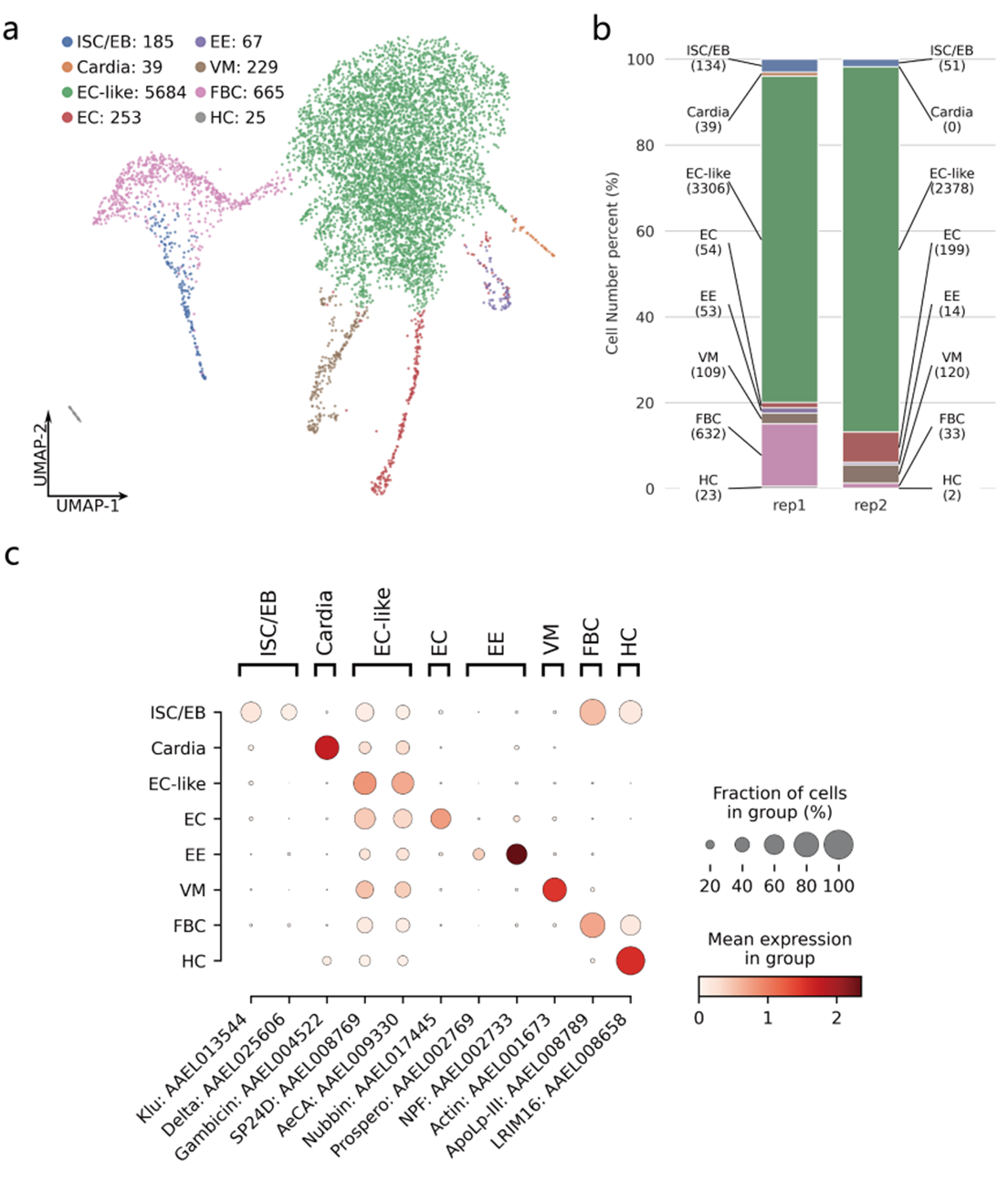
Fig 2. Cell type annotation for cell clusters (a) Annotation for different cell types by UMAP; (b) Percentage of the number of different cell types; (c) Marker gene for different cell types